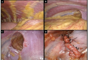
Thoracoscopy

To visually inspect the lungs, pleura, or mediastinum for evidence of abnormalities
To obtain tissue biopsies o fluid samples from the lungs, pleura, or mediastinum in order to diagnose infections, cancer, and other diseases
Used therapeutically to remove excess fluid in the pleural cavity or pleural cysts, or to remove a portion of diseased lung tissue (wedge resection).
To evaluate patients with pulmonary disease or abnormalities of the sac that surround the heart (pericardium) or the lining of the chest (pleura)
To obtain a tissue sample (biopsy) for further evaluation and to diagnose inflammation, infection, fibrosis and cancer
As a minimally-invasive method to perform certain types of surgery, such as pericardiectomy
Risks and Complications of Thoracoscopy
Thoracoscopy requires general anesthesia, and thus carries the associated risks.
Rare complications include excessive bleeding, infection, perforation of the diaphragm, and pneumothorax (leakage of air outside the lungs and into the pleural cavity, resulting in a collapsed lung).
After the Thoracoscopy
You will remain in the hospital for up to several days until you recover from the effects of surgery and anesthesia. During this time, your vital signs will be monitored, and you will be observed for any signs of complications.
You may be given pain-relieving medication to allay the discomfort associated with surgery.
A chest x-ray will be performed to ensure complete reinflation of the lung
Interstitial lung disease
Interstitial lung disease (ILD), or diffuse parenchymal lung disease (DPLD), is a group of lung diseases affecting the interstitium (the tissue and space around the air sacs of the lungs). It concerns alveolar epithelium, pulmonary capillary endothelium, basement membrane, perivascular and perilymphatic tissues.
Interstitial lung disease is a general category that includes many different lung conditions. All interstitial lung diseases affect the interstitium, a part of the lungs’ anatomic structure.
The interstitium is a lace-like network of tissue that extends throughout both lungs. The interstitium provides support to the lungs’ microscopic air sacs (alveoli). Tiny blood vessels travel through the interstitium, allowing gas exchange between blood and the air in the lungs. Normally, the interstitium is so thin it can’t be seen on chest X-rays or CT scans.
Causes of Interstitial Lung Disease
Bacteria, viruses, and fungi are known to cause interstitial pneumonia. Regular exposures to inhaled irritants at work or during hobbies can also cause some interstitial lung disease. These irritants include:
- Asbestos
- Silica dust
- Talc
- Coal dust, or various other metal dust from working in the mining
- Grain dust from farming
- Bird proteins (such as from exotic birds, chickens, or pigeons)
